I take the opportunity to thank you for choosing or showing interest in M. L. Jain Diagnocare. At M. L. Jain Diagnocare, it is our constant endeavor to provide you with an experience to cherish. You can enjoy a host of diagnostic services with care & quality. All our services are available from 7 am to 7 pm. In case of any query you can call our helpline number: 908 311 9991/9992 or communicate through email (mljaindiagnocare@gmail.com) or website www.mljaindiagnocare.com or through our Facebook page. We wish you a long and happy association with MLJD.
Warm regards,
Dr. Kaushik Saha.
Chief Pathologist.
Warm regards,
Dr. Kaushik Saha.
Chief Pathologist.
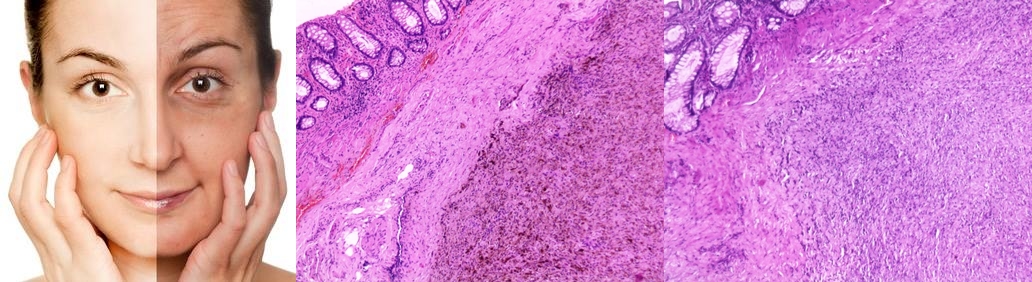 We are very much familiar with the term facial bleaching which dilutes or destroys or removes the concentrated melanin in the skin and helps in whitening the skin. Similarly we have to use bleaching agents for tissue sections especially in case of malignant tumor of melanocyte /melanoma where large amount of melanin obscures the cellular detail and negatively impacts immunohistochemical analysis of melanin-containing tissue samples by direct physical masking of antibody–antigen interactions. Today we have successfully removed the melanin pigments from tissue sections of a rectal melanoma. Thanks to MLJ Laboratory technicians.
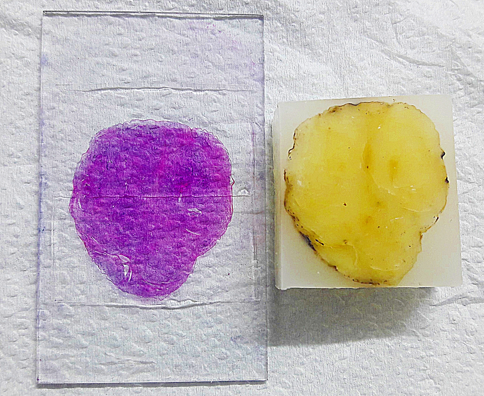 Large-Format Histopathology [LFH] preserves the spatial relationship of the tumor components and their relationship to the resection margins & helps to assess subgross morphological prognostic parameters like disease extent, lesion distribution, and tumor size. LFH is also used for the digitalization of glass slides in this new era of Whole Slide Imaging [WSI] where we can share the image for teaching, consultation, remote interpretation, and quality assurance. Usually, LFH is used in few developed countries especially for breast mass, prostate, brain tumor, intestinal polyp etc. But it is difficult to embed and process such a large tissue in one slide (45 × 75 mm). We are very proud to introduce LFH with the support of our genuinely expert technicians.
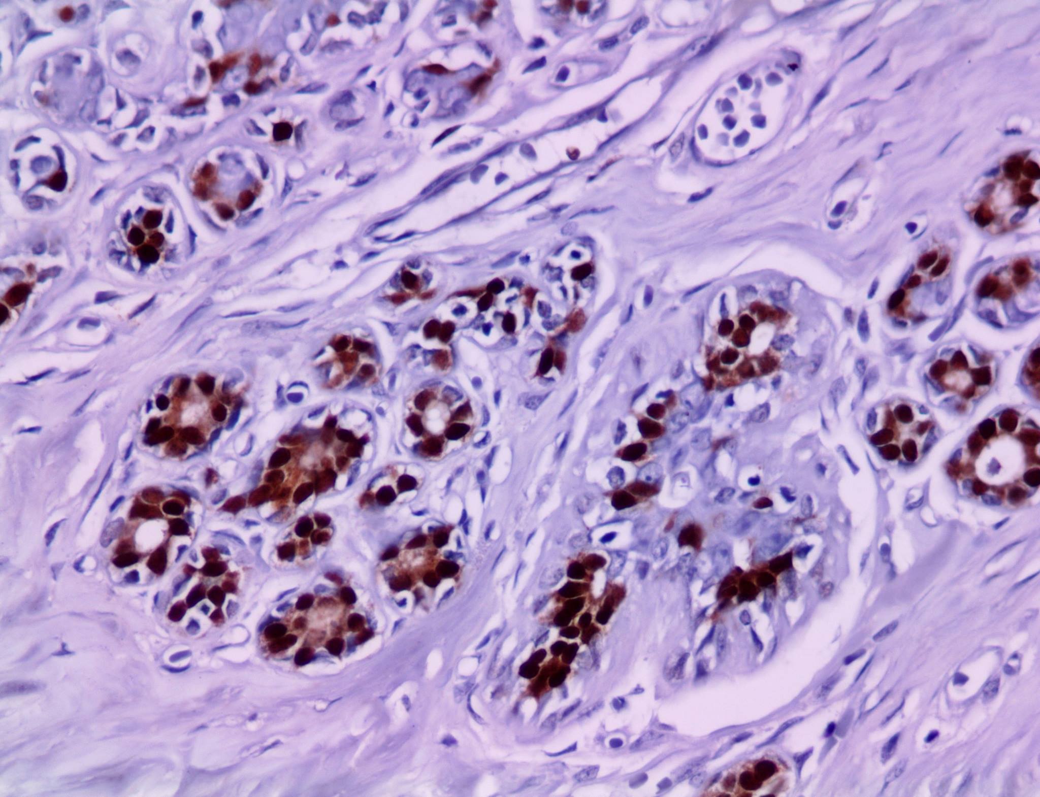 We have started another facility in tissue diagnosis -Immunohistochemistry (IHC), a method for localizing specific antigens in tissues or cells based on antigen-antibody recognition. At present only few centers in Kolkata are doing IHC routinely. It is an excellent method for tumor diagnosis especially for lymphoma, soft tissue tumor & metastatic tumor. It is also very useful to judge the appropriate patient for hormonal therapy like breast cancer or targeted therapy in lung cancer. 25.10.2017
 Today we have started another facility in tissue diagnosis - Cell Block Preparation, which is the bridge between histology & cytology, having advantages of future special staining, immunohistochemical or immunocytochemical staining, better architecture & pattern interpretation, long term storage & multiple sections as and when required. We will provide the paraffin block & stained slide along with FNAC slides on the next day of the procedure in clinically indicated cases.